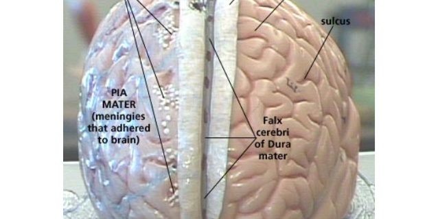
Our theme of the neural fascia continures this week with a look at the arachnoid mater, the second layer of protective fascial tissue around the brain and spinal cord, both beautifully demonstrated in these images supplied through “Wickiwand”. The arachnoid mater is a derivative of the neural crest mesectoderm in the embryo, and while it has no attachment to the more superficial Dura Mater, these two structures fuse at the level of S2 forming the filum terminale. Cerebrospinal fluid (CSF) flows beneath the arachnoid in the subarachnoid space, within a meshwork of trabeculae which connect the arachnoid and the deeper pia. The arachnoid mater makes arachnoid villi, tiny protrusions through the dura mater into the venous sinuses of the brain. This allows CSF to exit the subarachnoid space and enter the blood stream.
Interestingly, the arachnoid mater and the deeper pia mater are generally non-vascular. However, there is a network of veins that lie between the dura and arachnoid maters connecting the brain’s venous system with the venous system in the dura mater.
See you next week for a look at the Pia Mater, and in the meantime, #StayFasciannated.